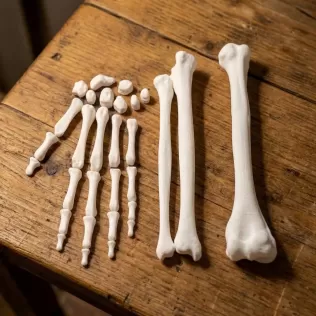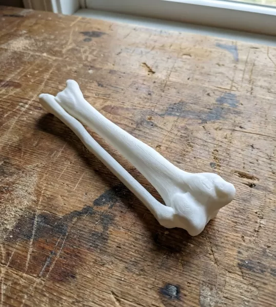
Radius & Ulna – 100% Anatomically Accurate – Asklepios Series
m
ID: 29544
$4.88
3D Model Details
-
Texture: No
-
Multipart model: No
-
File format: stl
-
File size: 3.42 MB
-
Upload date: 05/05/2026
-
Last update: 05/05/2026
Description
Are you looking for a study aid that isn't just a "visual approximation" but a faithful reflection of human biology? 🎓
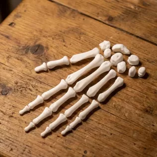
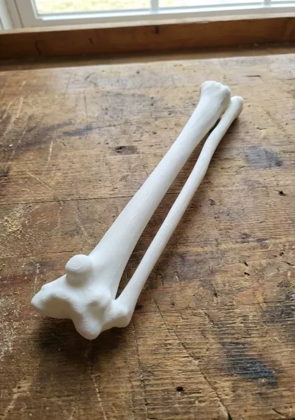
Introducing the Radius and Ulna (Forearm Bones) model, part of the prestigious Asklepios Series. This model was created using a high-resolution 3D scan (CT/DICOM) of an actual human specimen, making it an unrivaled resource for medical students, physiotherapists, and orthopedic professionals.
✅ What makes this model unique?
📍 100% Anatomical Fidelity: This is not an artistic sculpt. It is a precise digital twin of a human bone, capturing every tubercle, notch, and articular surface with micron-level accuracy.
🦴 Realistic Surface Texture: Because it is derived from a real scan, the print reveals natural bone irregularities, muscle attachment sites (apophyses), and nutrient foramina.
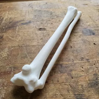
🔬 Educational Precision: Perfect for mastering osteology, practicing palpation, or understanding the complex mechanics of the radioulnar joints.
Key Anatomical Landmarks Included:
This model allows for a detailed study of structures such as:
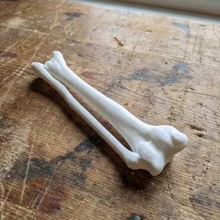
On the Radius: The Caput radii (radial head), Tuberositas radii (radial tuberosity), and the prominent Processus styloideus (styloid process).
On the Ulna: The Olecranon (elbow point), Incisura trochlearis (trochlear notch), Incisura radialis, and the Coronoid process.
The model perfectly depicts the interosseous space, crucial for understanding the biomechanics of pronation and supination.
Introducing the Radius and Ulna (Forearm Bones) model, part of the prestigious Asklepios Series. This model was created using a high-resolution 3D scan (CT/DICOM) of an actual human specimen, making it an unrivaled resource for medical students, physiotherapists, and orthopedic professionals.
✅ What makes this model unique?
📍 100% Anatomical Fidelity: This is not an artistic sculpt. It is a precise digital twin of a human bone, capturing every tubercle, notch, and articular surface with micron-level accuracy.
🦴 Realistic Surface Texture: Because it is derived from a real scan, the print reveals natural bone irregularities, muscle attachment sites (apophyses), and nutrient foramina.
🔬 Educational Precision: Perfect for mastering osteology, practicing palpation, or understanding the complex mechanics of the radioulnar joints.
Key Anatomical Landmarks Included:
This model allows for a detailed study of structures such as:
On the Radius: The Caput radii (radial head), Tuberositas radii (radial tuberosity), and the prominent Processus styloideus (styloid process).
On the Ulna: The Olecranon (elbow point), Incisura trochlearis (trochlear notch), Incisura radialis, and the Coronoid process.
The model perfectly depicts the interosseous space, crucial for understanding the biomechanics of pronation and supination.
Report this design